KPV Peptide in Canada: The Alpha-MSH Tripeptide, Explained
- Last reviewed
- Nov 9, 2026: every PMID listed here was opened and checked on this date.
- Reviewed by
- Peak Peptides Research Team
- Evidence labels
- In vitroAnimalHuman
- How we evaluated the evidence
- We read every cited abstract, and the open full text where it was available, labelled each finding by model, and quoted doses only when the abstract or open full text stated them.
- Disclosure
- Peak Peptides sells research compounds discussed here.
In short
KPV (Lys-Pro-Val) is the C-terminal tripeptide of alpha-MSH. In human intestinal cell lines, 10 nM KPV entered cells through PepT1 and reduced NF-κB activity.[1] Mouse colitis models used 100 μM KPV in drinking water. No published human trials were found. Researchers can review a KPV research peptide batch certificate.
- What is KPV?
- Why the fitness and wellness community is researching KPV
- How is KPV thought to work?
- Research timeline
- What do intestinal and colitis models show?
- What does skin research show?
- Oral, nanoparticle or topical? The delivery problem
- How does KPV compare with BPC-157 and TB-500?
- Why researchers study KPV alongside repair peptides (the KLOW stack)
- What doses were used in published research?
- How to read a KPV COA
- FAQ
- References
Key takeaways
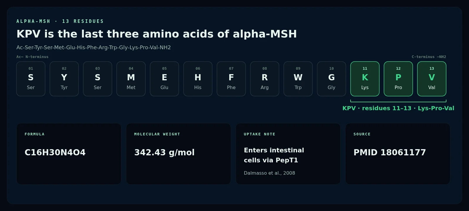
- A three-residue fragment. KPV is lysine-proline-valine, residues 11–13 of alpha-MSH (342.43 g/mol).[1]
- A transporter-dependent proposal. In intestinal and T-cell lines, nanomolar KPV inhibited NF-κB and MAP kinase signalling after PepT1 uptake.[1]
- Community interest is broader than the human evidence. Questions about gut, skin and “inflammation without tanning” map onto mouse and cell models, not onto trials in people.
- No published human trials of KPV were found. This guide therefore reports study design, not personal-use instructions, and there is no human dosing data.[1][2]
What is KPV?
KPV is a synthetic tripeptide used in laboratory research. It is the C-terminal fragment of alpha-melanocyte-stimulating hormone (alpha-MSH), a 13-residue peptide derived from proopiomelanocortin.
Its sequence is Lys-Pro-Val. Peak’s current certificate lists C16H30N4O4, 342.43 g/mol, CAS 67727-97-3 and H-Lys-Pro-Val-OH; the KPV research peptide page publishes that file. Reviews discuss alpha-MSH beyond pigmentation, including effects on immune cells and NF-κB.[6][7] The C-terminal fragment is α-MSH(11–13), or KPV; one review wrote that it “retains almost all of the anti-inflammatory capacity of the full hormone, but in its activities display a lack of any pigmentory action.”[2][5] That is why it appears in intestinal and skin-cell papers (see BPC-157 vs TB-500 vs KPV).
Why the fitness and wellness community is researching KPV
The fitness and wellness community is researching KPV for four overlapping reasons — gut health, skin, inflammation without tanning, and stacking it with other research peptides — each of which has a published laboratory counterpart and none of which has a human trial.
| Interest | Why people are interested | What the research shows | PMID |
|---|---|---|---|
| Gut health | PepT1, the transporter that carries KPV, is induced in inflamed colon in the IBD papers, so oral and colonic delivery are discussed as matching the site of injury. | PepT1-mediated uptake in intestinal and T-cell lines; mouse DSS, TNBS and T-cell-transfer colitis with lower myeloperoxidase activity or improved histology.[1][2] | 18061177, 18092346 |
| Skin | Alpha-MSH is a known cutaneous mediator, so readers ask whether its C-terminal fragment belongs in skin-cell research. | Calcium signals in human keratinocytes; a PM10 stress assay in HaCaT cells; a review of melanocortin peptides in wound research.[4][11][13] | 15102092, 40073467, 30661264 |
| Inflammation without tanning | Full alpha-MSH is known for pigmentary biology; the short fragment is discussed as a way to study inflammatory signalling without that activity. | Reviews state that KPV lacks the pigmentary action of the full hormone. A mouse peritonitis model separated its effect from core melanocortin-receptor signalling.[5][3] | 21222263, 12750433 |
| KLOW stacking | Research catalogues group KPV with BPC-157, TB-500 and GHK-Cu as a four-compound set. | No published study of that combination was found among the sources reviewed here. | None found |
“Interest” is the search language people use. The next columns separate that interest from what the papers measured. A result in mice or keratinocytes is not evidence of the same result in people.
Gut health. PepT1 is upregulated in inflamed colon in the IBD papers, which is why oral and colonic delivery appear so often.[1] Mouse colitis studies reported less weight loss, lower myeloperoxidase activity or improved histology.[2]
Skin. KPV triggered calcium signals in keratinocytes and changed PM10 markers at 50 μg/mL in HaCaT cells.[4][11] A 2019 review of melanocortin peptides in cutaneous wound research is a review, not a KPV trial.[13]
Inflammation without tanning. Several authors say the short fragment lacks the pigmentary activity of full alpha-MSH while still showing anti-inflammatory readouts in cells and mice.[5] Getting’s peritonitis model is the experimental illustration: KPV reduced cell accumulation by a route the authors judged different from core melanocortin peptides.[3]
KLOW stacking. The four-compound grouping is how KPV is often catalogued, not how it has been tested. No published combination study was found.
How is KPV thought to work?
The best-documented proposal is intracellular: PepT1 carries KPV into cells, after which NF-κB and MAP kinase signalling fall. Other work argues this activity is at least partly independent of the melanocortin-1 receptor.
PepT1 moves di- and tripeptides into cells. It is normally expressed in the small intestine and induced in the colon during inflammatory bowel disease. Dalmasso and colleagues found that Caco2-BBE, HT29-Cl.19A and Jurkat cells took up KPV through this transporter. Nanomolar KPV then inhibited NF-κB and MAP kinase activation; 10 nM lowered IL-1β-driven NF-κB reporter activity in Caco2-BBE cells. The paper reported a Km of about 160 μM in Caco2-BBE cells and about 700 μM in Jurkat cells — affinity in a dish, not a dose elsewhere.[1]
Full-length alpha-MSH often acts through MC1R. In two mouse colitis models KPV remained active when MC1R was non-functional; the authors concluded the effects “seem to be at least partially independent of MC1R signalling.”[2] In mouse peritonitis, KPV reduced neutrophil accumulation, was not blocked by an MC3/4 antagonist, did not raise macrophage cAMP, and was “unlikely to mediate its effects through melanocortin receptors.”[3] Reviews later attributed most of alpha-MSH’s reported anti-inflammatory activities to this tripeptide.[6][5] These proposals come from cells and mice, not from a human trial.
How has KPV research unfolded?
- 2000 Review of alpha-MSH as a cutaneous inflammatory modulator.[7]
- 2003 Mouse peritonitis: KPV separated from core melanocortin-receptor signalling.[3]
- 2004 Calcium signals recorded in human keratinocytes.[4]
- 2007 Review attributes most reported anti-inflammatory activities of alpha-MSH to KPV.[6]
- 2008 PepT1 uptake established; two mouse colitis models, including MC1R-deficient mice.[1][2]
- 2010 Colon-targeted nanoparticles; review notes the lack of pigmentary action.[8][5]
- 2017 HA-nanoparticles at 16 μg/kg/day in mice; permeation across microporated human skin.[9][12]
- 2019 Review of melanocortin peptides in cutaneous wound research.[13]
- 2021 Rectal KPV hydrogel in rat TNBS colitis.[10]
- 2025 50 μg/mL KPV in a HaCaT PM10 assay.[11]
What do intestinal and colitis models show?
The core in-vivo reports are mouse colitis studies from 2008. Formulation papers that delivered less peptide to a similar model are covered in the delivery section below.
In C57BL/6 mice, 100 μM KPV in drinking water for eight days with 3% DSS (five animals per group in the weight experiment) was associated with less weight loss, about 50% lower myeloperoxidase activity and less histologic damage; the same concentration was assessed 48 hours after TNBS.[1] Kannengiesser and colleagues reported earlier recovery in DSS and CD45RBhi transfer colitis and wrote that KPV “rescued all animals in the treatment group from death during DSS colitis” in MC1R-deficient mice. Their abstract does not state a dose.[2] These rodent readouts are not evidence from people and not instructions for use.
| Study | Year | Model | Finding (as reported) | PMID |
|---|---|---|---|---|
| Getting et al. | 2003 | Mouse crystal-induced and IL-1β peritonitis; MC1R-deficient mice | Reduced neutrophil accumulation; not blocked by an MC3/4 antagonist; still present in MC1R-deficient mice[3] | 12750433 |
| Elliott et al. | 2004 | HaCaT and normal human keratinocytes | Raised keratinocyte calcium at 10−15–10−7 M when cyclic AMP was inhibited; no cAMP rise detected[4] | 15102092 |
| Dalmasso et al. | 2008 | Caco2-BBE, HT29-Cl.19A, Jurkat; mouse DSS and TNBS colitis | PepT1 uptake; nanomolar NF-κB/MAPK inhibition; 100 μM in drinking water lowered colitis markers[1] | 18061177 |
| Kannengiesser et al. | 2008 | Mouse DSS and CD45RBhi transfer colitis; MC1R-deficient mice | Improved recovery and lower MPO; at least partly independent of MC1R[2] | 18092346 |
| Laroui et al. | 2010 | Caco2-BBE; mouse DSS colitis | Colon-targeted nanoparticles reduced inflammatory parameters; authors reported similar efficacy at a 12,000-fold lower concentration[8] | 19909746 |
| Xiao et al. | 2017 | FVB mouse DSS colitis; colonic epithelial cells and macrophages | HA-KPV-NPs at 16 μg/kg/day improved recovery versus non-coated particles[9] | 28143741 |
| Pawar et al. | 2017 | Dermatomed human skin | Iontophoresis plus microneedles raised permeation; passive diffusion was undetectable[12] | 28343991 |
| Sun et al. | 2021 | Rat TNBS colitis | Rectal hydrogel lowered colitis scores and myeloperoxidase; milligram amount not stated in the abstract[10] | 34547895 |
| Sung et al. | 2025 | HaCaT keratinocytes and a 3D skin model, PM10 exposure | 50 μg/mL restored viability and reduced IL-1β after PM10[11] | 40073467 |
Reviews by Luger, Brzoska and Böhm are cited in the text for interpretation, not as primary experiments.[5][6][7][13]
A clear limit
There are no published human trials of KPV in the sources reviewed here. The table reports cells, excised skin or rodents. Routes such as drinking water or hydrogel gavage are study design, not personal or veterinary methods.
What does skin research show?
Skin-directed papers ask whether KPV can signal in keratinocytes or change a pollutant-stress response in those cells. How it crosses skin is covered in the delivery section.
Elliott and colleagues did not detect a cyclic AMP rise after KPV in keratinocytes, but they recorded calcium responses at 10−15 to 10−7 M when PIA inhibited the cyclic AMP pathway.[4] Sung and colleagues reported that 50 μg/mL KPV restored HaCaT viability and reduced IL-1β after PM10 exposure.[11] Reviews place alpha-MSH among cutaneous mediators and ask whether shorter sequences belong in later wound research; they do not report a KPV trial.[7][13]
Oral, nanoparticle or topical? The delivery problem
Several groups asked whether a carrier could get KPV to intestinal tissue, or across skin, more efficiently than free peptide in solution. Those papers describe experimental design in animals or excised tissue, not a preferred route for people. For how published studies choose a route, see oral vs injectable peptides.
Laroui and colleagues loaded KPV into nanoparticles inside an alginate-chitosan hydrogel aimed at the colon, and reported similar efficacy in DSS-treated mice “at a concentration that is 12,000-fold lower than that of KPV in free solution.” The abstract does not give the two absolute amounts behind that ratio.[8] Xiao and colleagues used hyaluronic-acid-coated particles (about 272.3 nm) at 16 μg/kg of KPV per day as daily double hydrogel gavages for six days in FVB mice, and reported better weight recovery and lower TNF-α than with uncoated particles.[9] Sun and colleagues used a rectal KPV hydrogel in rat TNBS colitis and reported less weight loss and lower myeloperoxidase without stating the milligram amount.[10]
Pawar and colleagues asked a topical question. On dermatomed human skin, passive KPV permeation was below 0.01 μg/mL. Microneedles produced 4.4 μg/cm2/h, and iontophoresis plus microneedles raised that rate about 35-fold versus microneedles alone — a delivery experiment on excised skin, not a study of a skin condition.[12]
How does KPV compare with BPC-157 and TB-500?
The three peptides differ in length, origin and the models used to study them. Only BPC-157 and full-length thymosin beta-4 have any human reports in the sources cited here; KPV does not.
| Feature | KPV | BPC-157 | TB-500 |
|---|---|---|---|
| Identity | Lys-Pro-Val; alpha-MSH residues 11–13; 342.43 g/mol[1] | 15-residue gastric-juice fragment | Acetylated 7-residue fragment of thymosin beta-4 |
| Proposed mechanism | PepT1 uptake; NF-κB and MAP kinase inhibition; activity at least partly independent of MC1R[1][2] | NO, VEGFR2 and FAK-paxillin signalling in cell and rat studies | Inferred from parent Tβ4 G-actin binding |
| Main models | Human intestinal and skin cell lines; mouse and rat colitis models[1][2][10] | Rat gastric, vascular and tendon models | Parent Tβ4 in platelets and neutrophils; little fragment-specific animal work |
| Human data | No published trial found | Three small uncontrolled pilots (PMIDs 34324435, 39325560, 40131143)[15][16][17] | None found for the fragment. Full-length Tβ4 has, separately, a phase 2 dry-eye trial[14] |
Human findings for BPC-157 and thymosin beta-4 mark the contrast with KPV; they are not evidence for KPV. Sosne 2015 tested full-length thymosin beta-4, not the TB-500 fragment. See also BPC-157 vs TB-500 vs KPV and GHK-Cu copper peptide research.
Why researchers study KPV alongside repair peptides (the KLOW stack)
Researchers often discuss KPV beside BPC-157, TB-500 and GHK-Cu, a four-compound grouping supplied as the KLOW research stack, or beside BPC-157 alone as a gut research stack. Each compound has its own literature and a different proposed pathway. We found no published study that administered those combinations and measured an outcome, so stacking remains an open experimental question rather than an established protocol.
What doses were used in published research?
The figures below appear in the abstract or open full text of the papers we could open. They describe each study’s design in animals or cells. There is no human dosing data, because no human trials of KPV were found.
| Study | Year | Species / model | KPV form / delivery | Dose or concentration as reported | Duration | PMID |
|---|---|---|---|---|---|---|
| Elliott et al. | 2004 | HaCaT and normal human keratinocytes | Added to culture medium | KPV 10−15–10−7 M[4] | Acute calcium recordings | 15102092 |
| Dalmasso et al. | 2008 | Caco2-BBE, HT29-Cl.19A and Jurkat cells | Added to culture medium | 10 nM KPV[1] | Co-incubation with inflammatory stimuli (hours) | 18061177 |
| Dalmasso et al. | 2008 | C57BL/6 mouse DSS colitis (also TNBS) | Free peptide in drinking water | 100 μM KPV[1] | 8 days with DSS; colitis assessed 48 hours after TNBS | 18061177 |
| Xiao et al. | 2017 | FVB male mouse DSS colitis | HA-functionalized polymeric nanoparticles in a chitosan/alginate hydrogel, oral gavage | 16 μg/kg of KPV per day[9] | Daily double gavages for 6 days | 28143741 |
| Sung et al. | 2025 | Human HaCaT keratinocytes (PM10 assay) | Added to culture medium | 50 μg/mL KPV[11] | Not stated in the abstract | 40073467 |
These doses are reported from animal and cell studies for research reference only. They are not guidance for human or veterinary use. Kannengiesser 2008, Getting 2003, Laroui 2010, Sun 2021 and Pawar 2017 are omitted here because those sources did not state a usable KPV dose in the abstract or an open full text.
How to read a KPV COA
A certificate of analysis is the batch document that should travel with any research vial. Peak Peptides supplies KPV as a laboratory research material, not as a drug or health product.
On the certificate linked from the KPV research peptide page, check at least:
- Purity percentage. Peak’s specification is >98%.
- Batch or lot number. It should match the number on the vial.
- Test date.
- Named independent laboratory.
- Molecular weight. It should match 342.43 g/mol (C16H30N4O4). The current certificate also lists CAS 67727-97-3 and H-Lys-Pro-Val-OH.
Third-party tested, >98% purity, COA published. Ships from Toronto, discreet and tracked; same day if placed and paid before 12 PM Toronto time; 1–3 business days within Toronto, 3–5 business days Canada-wide. Payment by Interac e-Transfer.
Frequently asked questions about KPV
What is KPV peptide?
KPV is lysine-proline-valine, the last three amino acids of alpha-MSH. Peak’s certificate lists C16H30N4O4, 342.43 g/mol and CAS 67727-97-3. Published papers study it in intestinal cell lines, keratinocytes, mouse peritonitis and rodent colitis models. It is supplied here as a laboratory research material, not as a medicine.[1]
How is KPV thought to work?
The main proposal is that PepT1 carries KPV into intestinal and immune cells, after which NF-κB and MAP kinase signalling fall. Mouse work found activity at least partly independent of MC1R, and a peritonitis study argued that KPV does not act like core melanocortin agonists. These findings come from cells and animals, not from people.[1][2][3]
Are there human trials of KPV?
No published human trial of KPV was found in the sources reviewed for this article. The readouts summarised here come from cultured cells, excised human skin used as a permeation barrier, and mouse or rat colitis models. This guide therefore does not convert animal amounts into a human equivalent or offer personal-use directions.[1][9]
How does KPV compare with BPC-157?
They are different molecules. KPV is a three-residue alpha-MSH fragment studied mainly for PepT1-dependent NF-κB effects in gut and skin models. BPC-157 is a 15-residue peptide with three small uncontrolled human pilots. Those pilots are not KPV studies and do not close KPV’s human-evidence gap. A side-by-side comparison is in our earlier article.[1][15]
What should a KPV certificate of analysis include?
Look for a purity percentage (Peak’s specification is >98%), a batch or lot number that matches the vial, a test date, a named independent laboratory, and a molecular weight of 342.43 g/mol. Third-party tested, >98% purity, COA published. Compare the certificate on the product page with the vial you receive.
Is KPV good for gut health or skin?
That is the question many readers bring, and it is the wrong shape for the evidence. Mouse and rat colitis models, plus keratinocyte or excised-skin experiments, are why the fitness and wellness community discusses KPV. Those models do not establish a benefit in people. No published human trial has asked whether KPV is “good” for gut health or skin.[1][2][11]
Related research
BPC-157 vs TB-500 vs KPVThree mechanisms compared, labelled by model. oral vs injectable peptidesHow published studies choose a delivery route. GHK-Cu copper peptide researchA different tripeptide, with its own literature.Continue your research
Shop research-grade KPV at Peak Peptides
The KPV research peptide is supplied as lyophilized 10 mg reference material, with the current batch certificate published on the product page.
- Third-party tested, >98% purity, COA published.
- Ships from Toronto, discreet and tracked. Same day if placed and paid before 12 PM Toronto time; 1–3 business days within Toronto, 3–5 business days Canada-wide.
- Payment by Interac e-Transfer.
- For research use only. Not for human consumption.
Compounds in this article
Third-party tested · COA with every batch · shipped from Canada · for research use only
Get the Peak Peptides Research Letter
One email a week: the newest peptide studies, what they found, and the regulatory news that matters in Canada. Free, and you can unsubscribe anytime.
Subscribe freeReferences
- Dalmasso G, Charrier-Hisamuddin L, Nguyen HT, et al. PepT1-mediated tripeptide KPV uptake reduces intestinal inflammation. Gastroenterology. 2008;134(1):166-178. PMID 18061177 (PMC2431115) · PMID 18061177PMC2431115DOI 10.1053/j.gastro.2007.10.026 ↩
- Kannengiesser K, Maaser C, Heidemann J, et al. Melanocortin-derived tripeptide KPV has anti-inflammatory potential in murine models of inflammatory bowel disease. Inflamm Bowel Dis. 2008;14(3):324-331. PMID 18092346 · PMID 18092346DOI 10.1002/ibd.20334 ↩
- Getting SJ, Schiöth HB, Perretti M. Dissection of the anti-inflammatory effect of the core and C-terminal (KPV) alpha-melanocyte-stimulating hormone peptides. J Pharmacol Exp Ther. 2003;306(2):631-637. PMID 12750433 · PMID 12750433DOI 10.1124/jpet.103.051623 ↩
- Elliott RJ, Szabo M, Wagner MJ, et al. alpha-Melanocyte-stimulating hormone, MSH 11-13 KPV and adrenocorticotropic hormone signalling in human keratinocyte cells. J Invest Dermatol. 2004;122(4):1010-1019. PMID 15102092 · PMID 15102092DOI 10.1111/j.0022-202X.2004.22404.x ↩
- Brzoska T, Böhm M, Lügering A, et al. Terminal signal: anti-inflammatory effects of α-melanocyte-stimulating hormone related peptides beyond the pharmacophore. Adv Exp Med Biol. 2010;681:107-116. PMID 21222263 · PMID 21222263DOI 10.1007/978-1-4419-6354-3_8 ↩
- Luger TA, Brzoska T. alpha-MSH related peptides: a new class of anti-inflammatory and immunomodulating drugs. Ann Rheum Dis. 2007;66 Suppl 3:iii52-55. PMID 17934097 (PMC2095288) · PMID 17934097PMC2095288DOI 10.1136/ard.2007.079780 ↩
- Luger TA, Brzoska T, Scholzen TE, et al. The role of alpha-MSH as a modulator of cutaneous inflammation. Ann N Y Acad Sci. 2000;917:232-238. PMID 11268349 · PMID 11268349DOI 10.1111/j.1749-6632.2000.tb05388.x ↩
- Laroui H, Dalmasso G, Nguyen HT, et al. Drug-loaded nanoparticles targeted to the colon with polysaccharide hydrogel reduce colitis in a mouse model. Gastroenterology. 2010;138(3):843-853. PMID 19909746 · PMID 19909746DOI 10.1053/j.gastro.2009.11.003 ↩
- Xiao B, Xu Z, Viennois E, et al. Orally Targeted Delivery of Tripeptide KPV via Hyaluronic Acid-Functionalized Nanoparticles Efficiently Alleviates Ulcerative Colitis. Mol Ther. 2017;25(7):1628-1640. PMID 28143741 (PMC5498804) · PMID 28143741PMC5498804DOI 10.1016/j.ymthe.2016.11.020 ↩
- Sun J, Xue P, Liu J, et al. Self-Cross-Linked Hydrogel of Cysteamine-Grafted γ-Polyglutamic Acid Stabilized Tripeptide KPV for Alleviating TNBS-Induced Ulcerative Colitis in Rats. ACS Biomater Sci Eng. 2021;7(10):4859-4869. PMID 34547895 · PMID 34547895DOI 10.1021/acsbiomaterials.1c00792 ↩
- Sung J, Ju SY, Park S, et al. Lysine-Proline-Valine peptide mitigates fine dust-induced keratinocyte apoptosis and inflammation by regulating oxidative stress and modulating the MAPK/NF-κB pathway. Tissue Cell. 2025;95:102837. PMID 40073467 · PMID 40073467DOI 10.1016/j.tice.2025.102837 ↩
- Pawar K, Kolli CS, Rangari VK, et al. Transdermal Iontophoretic Delivery of Lysine-Proline-Valine (KPV) Peptide Across Microporated Human Skin. J Pharm Sci. 2017;106(7):1814-1820. PMID 28343991 · PMID 28343991DOI 10.1016/j.xphs.2017.03.017 ↩
- Böhm M, Luger T. Are melanocortin peptides future therapeutics for cutaneous wound healing? Exp Dermatol. 2019;28(3):219-224. PMID 30661264 · PMID 30661264DOI 10.1111/exd.13887 ↩
- Sosne G, Dunn SP, Kim C. Thymosin β4 significantly improves signs and symptoms of severe dry eye in a phase 2 randomized trial. Cornea. 2015;34(5):491-496. PMID 25826322 · PMID 25826322DOI 10.1097/ICO.0000000000000379 ↩
- Lee E, Padgett B. Intra-articular injection of BPC 157 for multiple types of knee pain. Altern Ther Health Med. 2021;27(4):8-13. PMID 34324435 Retrospective, uncontrolled. ↩
- Lee E, Walker C, Ayadi B. Effect of BPC-157 on symptoms in patients with interstitial cystitis: a pilot study. Altern Ther Health Med. 2024;30(10):12-17. PMID 39325560 Uncontrolled pilot. ↩
- Lee E, Burgess K. Safety of intravenous infusion of BPC157 in humans: a pilot study. Altern Ther Health Med. 2025;31(5):20-24. PMID 40131143 Two participants. ↩
Third-party tested, >98% purity, COA published. Ships from Toronto, discreet and tracked. Orders placed and paid before 12 PM Toronto time ship the same day; 1–3 business days within Toronto, 3–5 business days Canada-wide. Payment by Interac e-Transfer.
For research use only. Not for human consumption. This article summarises published research for educational purposes. It is not medical advice, not instructions for use and not a claim of benefit.